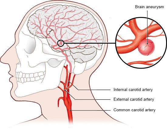
A brain aneurysm is a frail, swelling region in a corridor A
thick-walled vein conveying blood stream from the heart to any organ of the
body, including the brain. In the brain, closely resembling a dainty inflatable
or a shaky area on a tire's inner tube. Since its dividers might be week and
thin, an aneurysm is in danger of breaking. Assuming an aneurysm bursts, blood
spills into the space between the skull and the brain, a genuine kind of stroke
inability brought about by injury to the brain. Most strokes are brought about
by loss of blood stream to a piece of the brain (called an ischemic stroke or
cerebral localized necrosis) or by injury identified with draining inside the
brain tissue (an intracerebral hemorrhage) or into the space around the brain
(subarachnoid hemorrhage) known as a subarachnoid hemorrhage (SAH)
Types of brain aneurysms
Saccular
aneurysms, likewise called "berry" aneurysms since they seem as
though berries, are the most widely recognized kind of brain aneurysm. Saccular
aneurysms have a "neck" that interfaces the aneurysm to its primary
("parent") course and a bigger, adjusted region called the vault.
These aneurysms swell on just one side of the corridor divider. A more uncommon
sort is a fusiform aneurysm An sporadic molded extending of a cerebral vessel
that doesn't have a discrete neck or pocket. In which the conduit is extended
on the both sides. Fusiform aneurysms don't have a characterized neck.
Understanding the Brain
To understand aneurysms, it is useful to comprehend the
circulatory system of the brain. The heart pumps oxygen-and supplement loaded
blood to the brain, face, and scalp through two significant arrangements of
vessels: the internal carotid conduits and the vertebral courses. The throat
and different veins free blood once again from the brain
Warning Signs/Symptoms
Ruptured brain aneurysms ordinarily cause bleeding into the space around the brain, called a
subarachnoid hemorrhage.(SAH)Bleeding into the space around the brain (the
subarachnoid space)., which can cause sudden symptoms. Assuming you experience
any of the accompanying manifestations of a cracked aneurysm, CALL 911. It is
vital to comprehend that not these symptoms might be available; the best not
many recorded underneath are the most widely recognized.
Do not have a family member/companion take you in a private
vehicle to the emergency clinic. This is a high-stress circumstance that might
require specialists on call for use lifesaving methodology in the crisis
vehicle, and where time might be of the essence.
Sudden and severe headache, often described as “the worst headache
of my life”
1. Nausea/vomiting
2. Stiff neck
3. Blurred or double vision
4. Sensitivity to light
5. Seizure
6. Drooping eyelid
7. A dilated pupil
8. Pain above and behind the eye
9. Loss of consciousness
10. Confusion
11. Weakness and/or numbness
Unruptured
brain aneurysms usually have no symptoms. Typically, these aneurysms are
small. Many unruptured aneurysms are found incidentally when tests are being
done to screen for other conditions.
Rarely, unruptured aneurysms may become large and press on nerves
in the brain, causing symptoms. If you experience these symptoms, seek prompt
medical attention.
· 1. Blurred or double vision
· 2. A drooping eyelid
· 3. A dilated pupil
· 4. Pain above and behind one eye
· 5. Weakness and/or numbness
Unruptured aneurysms rarely cause chronic headaches, however acute
change in chronic headache pattern with respect to intensity or frequency would
be a good reason to reach out to your health care provider.
Causes/Risk Factors
Brain aneurysms grow quietly. Certain individuals might have
inherited a tendency for weak blood vessels, which might prompt the advancement
of aneurysms. Aneurysms in youngsters are uncommon, and most aneurysms likely
create because of mileage on the corridors all through an individual's
lifetime. Sometimes, extreme head injury or contamination might prompt the
improvement of an aneurysm.
There are various risk factors that add to the development of
aneurysms, listed below. Two of the most huge are, fortunately, ones that can
be controlled: cigarette smoking and
high blood pressure (hypertension).
· 1.Smoking
· 2. High blood pressure (hypertension)
· 3. Strong family history of brain aneurysms (familial
aneurysms)
· 4. Age (over 40)
· 5. Gender: women have an increased risk of
aneurysms
· 6. Race: people of color have an increased risk
of ruptured aneurysms
· 7. Other disorders: Ehlers-Danlos syndrome,
autosomal dominant polycystic kidney disease, Marfan syndrome, and fibro
muscular dysplasia
· 8. Presence of an arteriovenous malformation
(AVM)A particular type of vascular malformation of the brain. An abnormal
collection or tangle of arteries and veins located within the substance of the
brain in which a maldevelopment of capillaries (which normally connect the
arteries and veins) allows a high flow short cut through the brain.
· 9. Congenital abnormality in the artery A
thick-walled blood vessel carrying blood flow from the heart to any organ of
the body, including the brain. wall
· 10. Drug use, particularly cocaine
· 11 Excessive alcohol use
· 12. Infection
· 13 Severe head trauma
Diagnosis and Screening
Through imaging screening techniques, individuals at high risk of
harboring a brain aneurysm can be identified easily with non-invasive imaging
tests. An aneurysm is often diagnosed using a variety of imaging equipment.
Some methods include CT scan, CTA (computerized tomography angiography) -
In this procedure, a contrast dye is injected into the bloodstream prior to CT
scanning. This process produces detailed images of blood flow in the brain’s arteries. MRI
Short for magnetic resonance imaging. MRI is a painless, non-invasive procedure
that uses radio waves and a powerful magnetic field to produce detailed images
of the brain and other parts of the body. And MRA Short for magnetic
resonance angiography. MRA is a painless, non-invasive procedure that uses
radio waves and a powerful magnetic field to produce detailed images of blood
vessels. Sometimes an injected contrast dye is used..
Diagnosis
When a ruptured aneurysm is suspected, a head CT (computerized
tomography) scan is performed. This is a painless, non-invasive X-ray exam. A
CT scan will show if there has been bleeding in the brain.
However, a basic CT scan does not usually show the cause of the
bleeding. Using a technique called computerized tomography angiography (CTA),
in which a contrast dye is injected into the bloodstream, the brain’s blood
vessels are highlighted and aneurysms can be seen using special imaging
techniques.
Screening: Familial Aneurysms
In most cases, brain aneurysms are not hereditary, and there is
generally only a single case in a family. Occasionally, however, an individual
with a brain aneurysm will have other family members who are affected. When two
or more first-degree relatives (parent, child, or sibling) have proven
aneurysms, these are called “familial aneurysms.”
Individuals in these families may be at higher risk of developing
aneurysms than the general population. Therefore aneurysm screening with an
imaging study of the brain arteries is usually recommended, particularly for
first-degree relatives.
Neurologist
Follow
my social pages:
Facebook
https://facebook.com/Drjmohankrishna
Twitter:
https://twitter.com/drjmohankrishna
Instagram
https://instagram.com/drjmohankrishna
Linkedin
https://www.linkedin.com/mwlite/in/dr-j-mohan-krishna
pinterest
https://in.pinterest.com/Drjmohankrishna/
#drmohankrishna
#bestneurologist #Hyderabad #neurologist #strokedoctor #autoimmuneneurology
#besttreatment #NeurologyProblems #Prevention #brainhealth #neuroprevention #neuroproblems #neurohealth
#stroke
Follow
more Visit:

00 Reviews